Introduction:
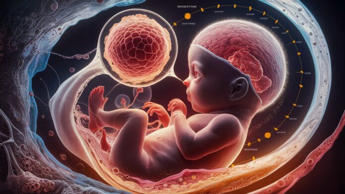
The journey of human development begins not at birth, but within the hidden sanctuary of the womb. Fetal life represents one of the most profound and complex biological processes, a meticulously orchestrated sequence of events transforming a single fertilized cell into a fully formed human being poised for independent existence. This period, spanning approximately thirty-eight weeks from conception to birth, is characterized by astonishingly rapid growth, intricate differentiation of tissues and organs, and the foundational establishment of bodily systems crucial for survival outside the uterine environment. Understanding this remarkable voyage illuminates not only the marvels of human biology but also underscores the critical importance of maternal health, prenatal care, and environmental influences on shaping an individual’s lifelong potential. This article delves deep into the captivating stages, key developmental milestones, influential factors, and essential care practices defining the extraordinary saga of fetal development.
The Foundational Stages of Prenatal Development
The odyssey of fetal life is formally divided into three primary trimesters, each marked by distinct developmental priorities and critical milestones. The first trimester, encompassing the initial twelve weeks, is arguably the most dynamic and vulnerable period. It commences with conception, the fusion of sperm and egg to form a zygote, which undergoes rapid cell division (cleavage) as it travels down the fallopian tube. Upon reaching the uterus, the developing blastocyst must successfully implant into the nutrient-rich uterine lining (endometrium), establishing the vital connection for future nourishment. This trimester witnesses the miraculous transformation from a cluster of cells into an embryo with rudimentary structures. Organogenesis, the formation of major organs and systems, occurs at a breathtaking pace. The neural tube closes, forming the foundation of the brain and spinal cord; the primitive heart begins to beat and pump blood; limb buds emerge; and the basic structures of the eyes, ears, and digestive system take shape. By the end of this phase, the embryo transitions into a fetus, signifying the completion of major structural formation.
Critical Processes Shaping Fetal Growth and Maturation
Following the groundwork laid in the first trimester, the second trimester (weeks 13-26) shifts focus towards tremendous growth and the refinement of existing structures. The fetus undergoes a dramatic increase in size and weight. Bones begin to ossify, transforming from cartilage to harder bone. Muscles develop and strengthen, enabling increasingly coordinated movements – a phenomenon perceived by the mother as quickening. Facial features become more defined, and external genitalia develop sufficiently for sex determination via ultrasound. Sensory systems advance significantly: the fetus develops the ability to hear sounds from the outside world, including the mother’s voice and heartbeat, and taste buds form. The third trimester (weeks 27 to birth) is dominated by maturation and preparation for extrauterine life. The primary objective is achieving viability and functionality of essential systems. The lungs undergo crucial development, producing surfactant – a substance critical for breathing air by preventing lung collapse. The brain experiences exponential growth and complexification, developing intricate folds (gyri and sulci) and establishing vital neural pathways. The fetus rapidly accumulates body fat, providing insulation and energy reserves. Reflexes, such as sucking and swallowing, become more coordinated. Simultaneously, the fetus typically assumes a head-down position (vertex presentation) in preparation for the journey through the birth canal.
Influential Factors and the Imperative of Prenatal Care
The trajectory of fetal development is profoundly influenced by a multitude of factors, highlighting the paramount importance of comprehensive prenatal care. Maternal health stands as the cornerstone. Chronic maternal conditions like uncontrolled diabetes, hypertension, or autoimmune disorders can significantly impact placental function and fetal growth. Adequate maternal nutrition is non-negotiable; sufficient intake of folic acid prevents neural tube defects, while protein, iron, calcium, and essential vitamins support organ development and growth. Conversely, exposure to teratogens – harmful agents such as alcohol, tobacco, illicit drugs, certain medications, infections (like rubella or Zika virus), and high levels of environmental toxins – can cause devastating birth defects, growth restrictions, or developmental disabilities, particularly during critical periods of organogenesis. Regular prenatal visits are indispensable. These appointments allow healthcare providers to monitor fetal growth through fundal height measurements and ultrasounds, assess fetal heart rate, screen for potential complications (like gestational diabetes or preeclampsia), provide essential vaccinations, and offer crucial guidance on nutrition, lifestyle, and risk avoidance. Advanced techniques like maternal-fetal medicine specialize in managing high-risk pregnancies and complex fetal conditions, utilizing sophisticated diagnostic tools like detailed anatomy scans, amniocentesis, or chorionic villus sampling (CVS).
The Vital Lifelines: Placenta, Umbilical Cord, and Amniotic Fluid
Sustaining fetal life hinges entirely on the flawless function of specialized supporting structures. The placenta, often termed the lifeline, is a unique organ formed jointly by maternal and fetal tissues. It acts as a sophisticated interface, performing the critical functions of nutrient transfer (delivering oxygen, glucose, amino acids, vitamins, and minerals from the mother’s bloodstream to the fetus), waste elimination (removing carbon dioxide and metabolic byproducts like urea from the fetal blood), gas exchange (facilitating the uptake of oxygen and release of carbon dioxide), and hormone production (generating hormones like progesterone, estrogen, and human chorionic gonadotropin (hCG) essential for maintaining pregnancy). Connected to the placenta is the umbilical cord, a flexible conduit containing one vein (carrying oxygenated, nutrient-rich blood to the fetus) and two arteries (returning deoxygenated, waste-laden blood to the placenta). Encasing the developing fetus is the amniotic sac, filled with amniotic fluid. This remarkable fluid serves multiple vital roles: it cushions the fetus against physical shocks and injuries, maintains a stable thermal environment, allows for freedom of movement crucial for musculoskeletal development, prevents the amniotic membranes from adhering to the fetus, possesses antimicrobial properties, and facilitates the development of the fetal lungs and digestive system as the fetus swallows and inhales the fluid.
Conclusion:
Fetal life is an extraordinary testament to the resilience and intricacy of human development. From the invisible moment of conception through the dramatic transformations of each trimester, the journey within the womb lays the essential physical and neurological groundwork for a lifetime. Understanding the stages of growth, the critical processes of differentiation and maturation, and the profound impact of maternal health, nutrition, and environmental factors underscores a powerful responsibility. Access to consistent, high-quality prenatal care is not merely beneficial; it is fundamental to optimizing outcomes, identifying potential risks early, and providing the supportive environment necessary for the fetus to reach its full potential. As we marvel at the intricate dance of fetal development, we are reminded of the profound interconnectedness between maternal well-being and the nascent life it nurtures, emphasizing that the journey towards a healthy birth begins long before the first cry is heard.
Frequently Asked Questions (FAQs)
- Q: What exactly defines the “fetal” stage versus the “embryonic” stage?
A: The embryonic stage spans from conception (fertilization) through the end of the 8th week of pregnancy. This period is focused on organogenesis – the formation of all major organs and body systems from the basic layers of cells. It’s a highly sensitive period for potential birth defects if exposed to teratogens. The fetal stage begins at week 9 and continues until birth. During this phase, the emphasis shifts from the initial formation of structures to tremendous growth, refinement, maturation of organs and systems, and the development of recognizable human features. The developing human is referred to as an embryo during the embryonic stage and a fetus during the fetal stage. - Q: When can the mother typically feel the baby move (“quickening”)?
A: The perception of fetal movements, known as quickening, is usually first noticed by the mother between 16 and 25 weeks of pregnancy, most commonly around 18-20 weeks for first-time mothers and potentially slightly earlier (16-18 weeks) in subsequent pregnancies. Early movements might feel like flutters, bubbles, or gentle taps. The frequency and strength of movements increase significantly as the fetus grows larger and stronger throughout the second trimester and into the third trimester. - Q: Why is folic acid so crucial before and during early pregnancy?
A: Folic acid (vitamin B9) is critically important in the very early stages of fetal development, particularly before conception and during the first few weeks (often before a woman knows she’s pregnant). It plays a vital role in the proper formation and closure of the neural tube, which develops into the baby’s brain and spinal cord. Adequate folic acid intake (typically 400-800 micrograms daily as recommended) significantly reduces the risk of serious neural tube defects (NTDs) like spina bifida and anencephaly. Since the neural tube closes very early (by around day 28 post-conception), ensuring sufficient levels before pregnancy begins is essential. - Q: What does “viability” mean in the context of fetal development?
A: Viability refers to the point in pregnancy where a fetus has developed sufficiently to have a chance of surviving outside the mother’s womb, albeit with significant medical intervention in most cases. This point is not fixed but has advanced due to improvements in neonatal intensive care. Currently, viability is generally considered possible around 23-24 weeks of gestation, though survival rates at this stage are still relatively low and associated with high risks of severe complications. Survival rates improve dramatically with each additional week spent in the womb. Viability depends heavily on the maturation of the lungs (ability to breathe air) and other vital systems. - Q: How does maternal stress impact fetal life?
A: While the placenta provides some protection, chronic, severe maternal stress can potentially impact fetal development and long-term outcomes. High levels of stress hormones (like cortisol) can cross the placenta. Research suggests potential links to effects such as increased risk of preterm birth, lower birth weight, altered fetal neurodevelopment, and potentially even influencing the baby’s stress response system later in life. While everyday stress is generally not harmful, managing significant, ongoing stress through healthy coping mechanisms (exercise, relaxation techniques, therapy, strong social support) is an important aspect of prenatal care for both maternal and fetal wellbeing.

